What is a Veterinary Ultrasound?
An ultrasound is a safe, non-invasive, and painless diagnostic tool that uses high-frequency sound waves to create real-time images of your pet’s internal organs. Unlike X-rays, which show bones and air-filled structures, ultrasounds allow us to see the texture, shape, and movement of soft tissues.
The Benefits: Why Choose Ultrasound?
When your pet isn’t feeling their best, we want answers fast. Ultrasound provides a “window” into their body without the need for surgery.
-
Real-Time Visualization: We can watch the heart beat, observe blood flow, and see organs functioning in real-time.
-
Early Detection: It is the gold standard for detecting tumors, cysts, or early-stage organ disease that might be missed by blood work and x-rays.
-
Stress-Free Procedures: Most pets remain calm during the process, often requiring little or no sedation.
-
Guided Precision: If a biopsy is needed, ultrasound allows us to guide the needle exactly where it needs to go.
-
No Radiation: Unlike X-rays, there is zero radiation exposure, making it safe for repeat exams and pregnant patients.
Ultrasound Procedures types we offer.
- Abdominal Ultrasound: The abdomen is home to the body’s most complex filtration and digestive systems. An abdominal ultrasound allows us to look “inside” organs like the liver, kidneys, and spleen to check for changes in size, texture, or the presence of masses. It is also an invaluable tool for identifying painful bladder stones or tracking down “foreign bodies”—like a swallowed toy or sock—that may be causing a life-threatening gastrointestinal disease and obstruction. By seeing the organ’s internal architecture, we can often catch chronic diseases, such as kidney failure or hepatitis, much earlier than with physical exams alone.
- Cardiac Imaging (Echocardiography): When it comes to the heart, motion is everything. An echocardiogram is a specialized ultrasound that allows us to watch the heart in action. We can measure the thickness of the heart walls, ensure the valves are opening and closing properly, and even use “Doppler” technology to see the direction and speed of blood flow. This is the primary way we diagnose heart murmurs, congenital defects, or congestive heart failure, allowing us to tailor a medication plan that can significantly extend a pet’s quality of life.
- Pregnancy & Reproductive Health: Ultrasound is the safest and most exciting way to monitor a growing litter. Unlike X-rays, which are usually reserved for the very end of pregnancy to count bones, ultrasound can confirm pregnancy as early as 20–25 days after breeding. It allows us to visualize the gestational sacs and, most importantly, monitor fetal heartbeats to ensure the puppies or kittens are healthy and thriving. Beyond pregnancy, it is also used to screen for “pyometra”—a severe, life-threatening uterine infection—providing a quick diagnosis when time is of the essence.
- Emergency (TFAST/AFAST) Scans: In an emergency, every second counts. We use rapid ultrasound protocols (known as TFAST or AFAST) to perform a “quick look” at the chest and abdomen of a patient who has experienced trauma, such as a fall or a car accident. These scans are designed to find “free fluid” (internal bleeding) or air trapped outside the lungs. Because these scans can be done while the pet is being stabilized, they provide the veterinary team with immediate, life-saving information without the need for the patient to be moved to a separate X-ray room.
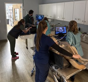
We strive to make the experience as relaxing as possible for your pet. Here is how the process usually goes:
- Most pets need to fast (no food) for 8-12 hours prior to an abdominal ultrasound to ensure the stomach is clear.
- In some instances, oral sedative medication will be dispensed prior to appointment.
- To get a clear image, sound waves need direct skin contact. We will gently shave a small patch of fur in the area being examined.
- Your pet will lay on a padded “V-trough” or a soft mat. A cool, water-based gel is applied to the skin, and a probe (transducer) is moved across the area.
- Most scans take between 20 to 45 minutes, depending on the complexity of the case.
- In many cases, we can discuss preliminary findings immediately. For complex cases, the images may be sent to a board-certified radiologist for a formal report.

